
Application Recommendation: The Use of Confocal Raman Microsope in Kidney Health
Introduction
Urinary calculi and kidney diseases are global issues, with crystallization being a significant diagnostic indicator of urological stones and kidney damage. The etiology of urolithiasis is complex and has a clear relationship with metabolic abnormalities. Analyzing the composition of urinary stones can help understand metabolic risk factors, playing an important role in analyzing the causes of stones, and preventing their formation and recurrence. Therefore, this study aims to identify and analyze urinary crystals based on Raman spectroscopy technology, which is used for the diagnosis of urological stones and kidney damage. The prevalence of urinary calculi and kidney diseases varies even within different regions of the same province. Studying the unchangeable factors such as gender, age, race, and genetics of regional urinary stone patients can provide effective evidence for local urological stone screening and early intervention treatment. Research on changeable factors such as environmental influences and dietary habits of regional urinary stone patients can provide theoretical basis for preventive measures against the recurrence of urinary stones and for local health guidelines.
Background
The incidence of urological stones in China is between 1% and 5%. In recent years, there have been significant advances in both basic research and clinical treatment of urological stones. Different urinary crystals have different clinical diagnostic significance. Traditional methods of identifying urinary crystals mainly relied on chemical reagents and microscopic observation, which are labor-intensive, resource-consuming, and do not protect the samples well. However, with the rapid development of Raman spectroscopy technology, it is now possible to quickly identify and classify urinary crystals.
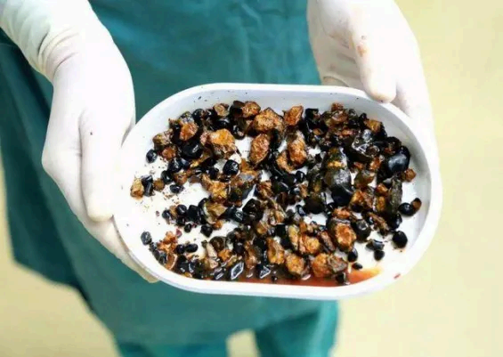
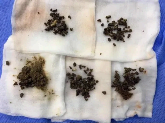
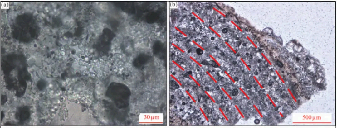
Stones Extracted During Surgery:
Instrument Introduction
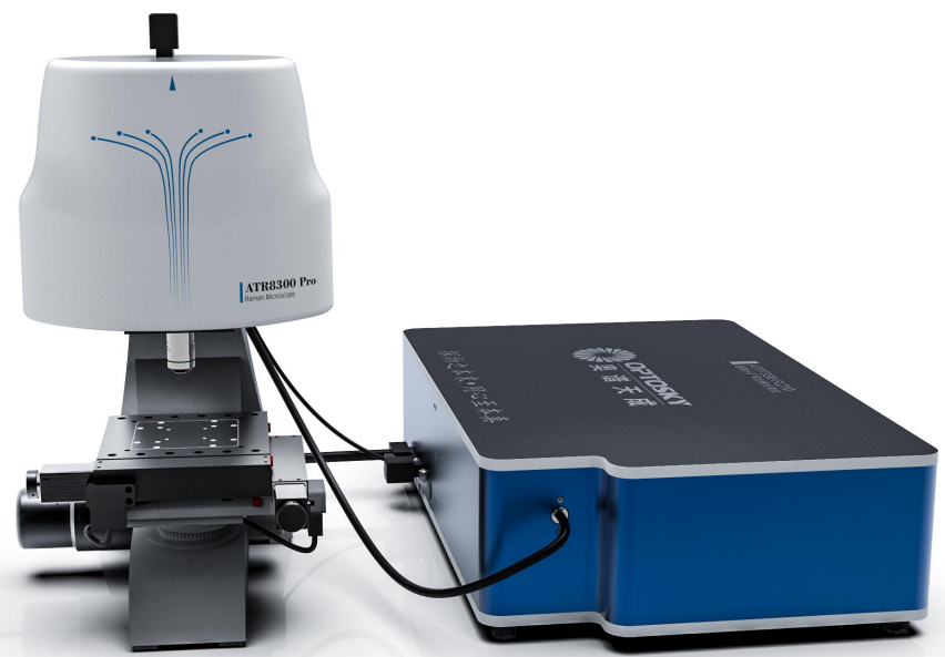
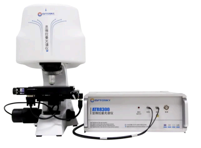
The ATR8300 is equipped with objectives specifically designed for Raman systems, allowing the laser spot to approach the diffraction limit. Through a 3-megapixel camera, the focus information is accurately and intuitively displayed on the computer, overcoming the common issue in ordinary Raman systems where the collection plane of the Raman signal is slightly above or below the actual optimal focal plane. This enhances the quality of Raman spectra.
The ATR8300 has no moving parts for optical path switching; all optical components are solid-state assembled, making the operation very stable. This setup perfectly solves the issue of light path loss during camera imaging and achieves the separation of camera imaging and Raman signal collection, thereby obtaining the best signal intensity.
Raman spectroscopy is a technique that reflects molecular vibrations and rotations, essentially a characteristic frequency shift generated by the interaction between molecules and photons that characterizes molecular vibration or rotational energy level differences. By analyzing the Raman frequency shift through Raman spectroscopy, structural and property analysis at the molecular level of a substance can be conducted. Raman spectroscopy allows for the determination of molecular structure and chemical properties, identifying specific molecular types or components, thereby enabling the rapid identification and analysis of urinary crystals.
|
Substance |
Raman Frequencies/cm⁻¹ |
|
Bird Guano Stone (Struvite) |
965、566 |
|
Carbonate Apatite |
970、1078 |
|
Calcium Oxalate Stone |
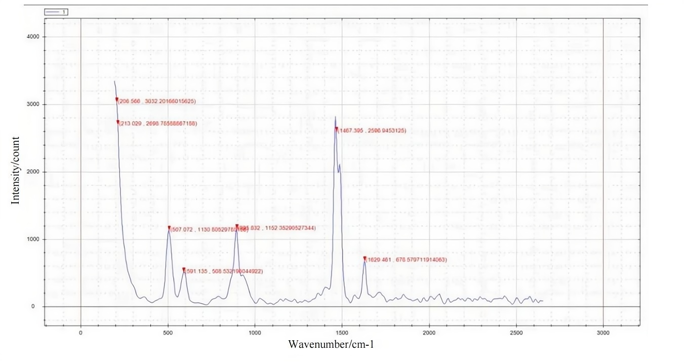
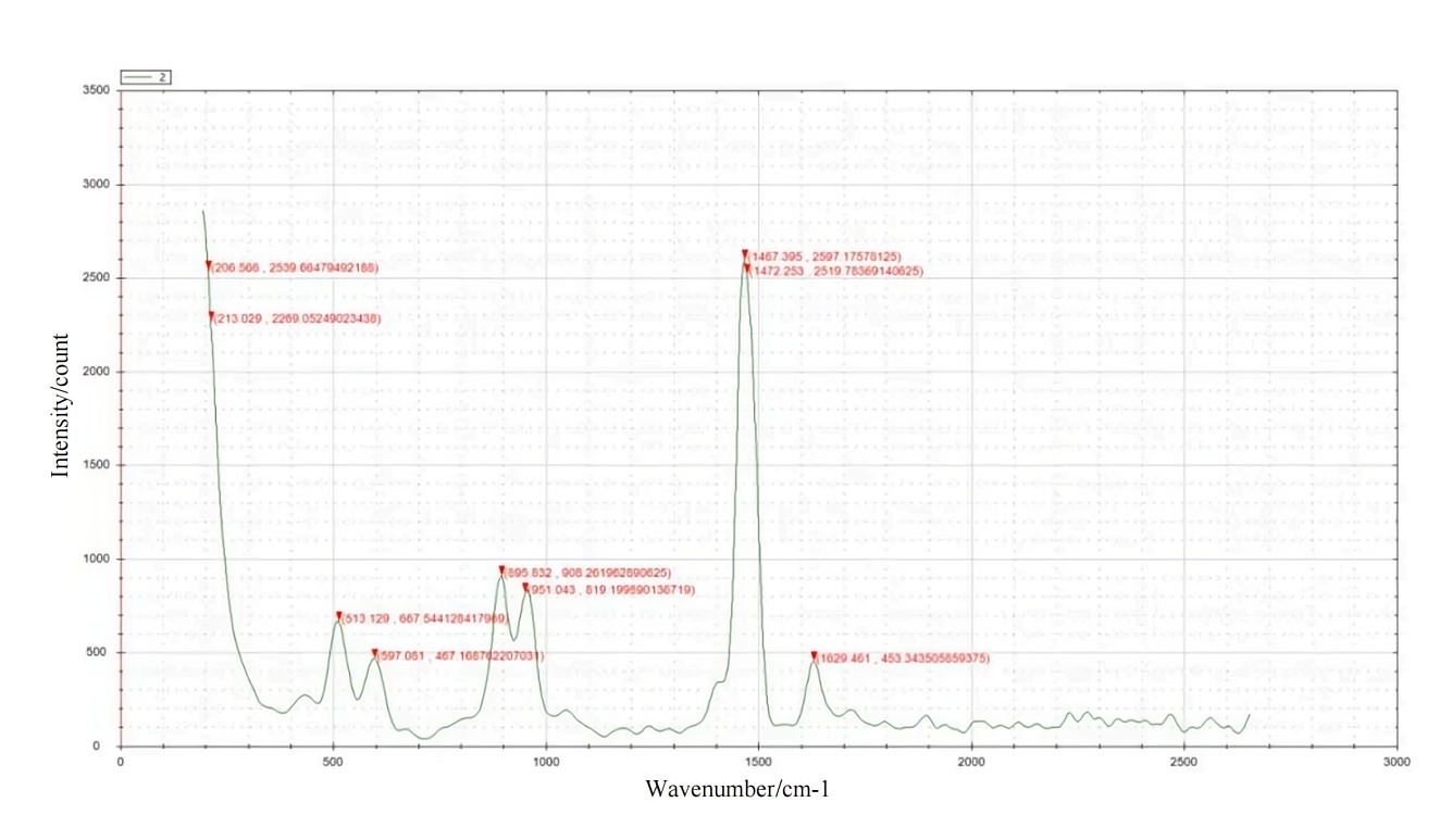
1474、910、597、1480、920、513、1467、1490、1630、895、951 |
Referring to previous Raman spectroscopy studies of urinary stones, the vibrational modes of calcium oxalate stones are more complex than those of phosphates, featuring multiple strong characteristic peaks. The frequencies of different vibrational modes on the Raman spectra primarily lie in the 400-1800 cm⁻¹ range, including symmetric stretching vibrations of carboxylate groups in the 1400-1550 cm⁻¹ range, asymmetric stretching vibrations in the 1550-1700 cm⁻¹ range, C-C symmetric stretching vibrations in the 850-950 cm⁻¹ range, and O-C-O in-plane bending vibration modes (β(O-C-O)) in the 450-550 cm⁻¹ range. Additionally, calcium oxalate stones show variations in their Raman spectra due to differences in water content, leading to molecular structural deviations. The Raman spectra of the common components of urinary stones, monohydrate calcium oxalate (whewellite) and dihydrate calcium oxalate (weddellite), show significant differences. Monohydrate calcium oxalate exhibits double peaks at 1467 cm⁻¹ and 1490 cm⁻¹, a peak at 1629 cm⁻¹, and a peak at 895 cm⁻¹ corresponding to C-C vibrations.
Product Recommendation|Application Case Study of ATR3000-Mobile Raman Spectrometer
Application Of Spectral Technology In Seed
Related Article