
Quantitative Determination Of Blood Glucose By Raman Spectroscopy
author: Joy
2022-01-07
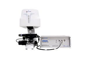
At present, blood glucose detection is generally carried out by blood collection. In short, a certain amount of blood is extracted and the concentration of glucose in the blood is detected by biological or physical methods. A few microliters of blood sample should be taken for the least damage, and the concentration of blood glucose is calibrated by enzymatic reaction. At present, nondestructive testing methods have not been widely used in clinic. The main problems are as follows: 1. The blood glucose concentration is very low, and the clinical accuracy cannot be accurately achieved by traditional physical means;2. Current lossless technical methods cannot obtain direct single information of blood due to skin or other interference, and generally extract relevant information through mathematical processing, resulting in large interference, unstable and inaccurate measurement; 3. There is no internal calibration reference index due to the large difference of individual skin resulting in large spectral difference.Blood glucose can be quantitatively determined by Raman spectroscopy.
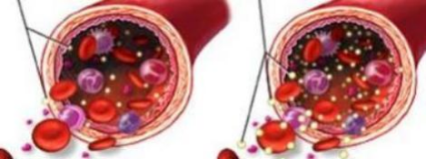
Principle: Raman Scattering Spectroscopy
Light on matter scatters both elastic and inelastic. The scattered light of elastic scattering has the same wavelength as the excited light, while the scattered light of inelastic scattering has the longer and shorter components than the excited light, collectively known as the Raman effect.The invention relates to a nondestructive method for quantitative detection of blood glucose using Raman spectroscopy, including the following steps;Laser light from a semiconductor diode, which uses a lens to pass through the skin, is focused into the blood vessels;To obtain the Raman spectrum of blood, through the Raman spectra obtained by Jane characteristic peaks of glucose and hemoglobin characteristic peak intensity division by glucose peak relative strength, calculate the ratio of different blood glucose concentration and glucose peak relative intensity was relatively strong and the concentration curve, the curve as the basis of quantitative measuring blood sugar levels;The relative intensity of glucose peak in Raman spectrum of blood with unknown concentration was compared with the relative intensity - concentration curve of blood glucose to obtain the glucose concentration in blood.
The laser penetrates the skin and focuses into the blood vessels to obtain the Raman spectrum.of blood. The characteristic peaks of glucose and hemoglobin are used to judge the concentration of blood glucose. Thecharacteristic peak of hemoglobin is used as the internal calibration index to overcome the interference of skin and other factors on the signal and the problem of signal size variation. Thus, accurate data of blood glucose concentration can be obtained.The method of nondestructive measurement of blood glucose in skin by Raman spectroscopy includes the following steps: 1) A laser beam from a semiconductor diode is introduced into a lens through an interference filter, and then focused under the skin;
2) Adjust the height of focus so that the focus is in the blood. The height adjustment can be controlled by piezoelectric ceramics or by moving the skin, and the determination of the appropriate focus can be achieved by Raman spectroscopy. If there is a relatively single blood spectrum, it is considered that the location is appropriate;
3) The Raman spectral signal excited by the focus is introduced into the spectrometer after passing through the notch filter or band pass filter. The spectrometer is connected with the CCD detector, and the Raman spectral information such as blood is obtained by THE CCD.
4) Extract the characteristic peaks of glucose and hemoglobin from the spectral data;
5) The relative intensity of glucose peak was obtained by dividing the intensity of glucose characteristic peak (1125cm') and hemoglobin characteristic peak (1549cm') obtained by Raman spectroscopy;
The laser penetrates the skin and focuses into the blood vessels to obtain the Raman spectrum.of blood. The characteristic peaks of glucose and hemoglobin are used to judge the concentration of blood glucose. Thecharacteristic peak of hemoglobin is used as the internal calibration index to overcome the interference of skin and other factors on the signal and the problem of signal size variation. Thus, accurate data of blood glucose concentration can be obtained.The method of nondestructive measurement of blood glucose in skin by Raman spectroscopy includes the following steps: 1) A laser beam from a semiconductor diode is introduced into a lens through an interference filter, and then focused under the skin;
2) Adjust the height of focus so that the focus is in the blood. The height adjustment can be controlled by piezoelectric ceramics or by moving the skin, and the determination of the appropriate focus can be achieved by Raman spectroscopy. If there is a relatively single blood spectrum, it is considered that the location is appropriate;
3) The Raman spectral signal excited by the focus is introduced into the spectrometer after passing through the notch filter or band pass filter. The spectrometer is connected with the CCD detector, and the Raman spectral information such as blood is obtained by THE CCD.
4) Extract the characteristic peaks of glucose and hemoglobin from the spectral data;
5) The relative intensity of glucose peak was obtained by dividing the intensity of glucose characteristic peak (1125cm') and hemoglobin characteristic peak (1549cm') obtained by Raman spectroscopy;
6)The relative strength concentration relationship curve was obtained by calculating the relative intensity of different glucose concentrations and
glucose peaks, which was used as the basis for quantitative measurement of glucose concentrations;
glucose peaks, which was used as the basis for quantitative measurement of glucose concentrations;
7) The relative intensity of glucose peak in the blood Raman spectrum of unknown concentration was compared with the relative intensity - concentration curve to obtain the glucose concentration in the blood.
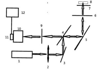
Fig 1System Optical Path Structure Diagram.In FIG. 1, 1. Semiconductor laser diode, 2. Interference filter, 3. Bicolor mirror, 5. Reflector, 6. Lens, 7. Slide, 8. Blood vessel, 9. Band pass or holographic filter, 10. Spectrometers, 11.CCD, 12. Computer.
Result:
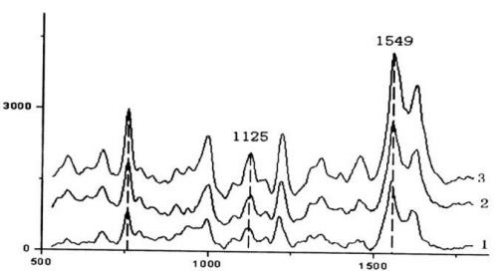
Fig 2Raman Spectra OfDifferent Blood Glucose Concentrations Obtained From Blood Vessels Under The Skin
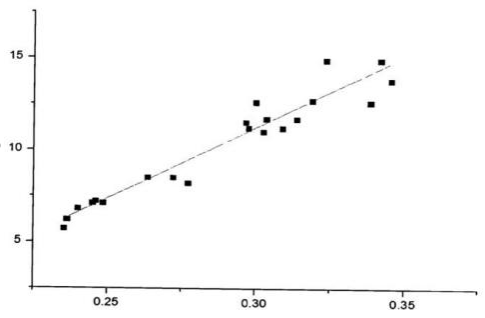
Fig 3The Relative Strength - Concentration Curve Of Glucose.
Conclusion:
Raman spectroscopy can quantitatively determine blood glucose, which is a nondestructive testing method, convenient and quick.Using OPTOSKY’S ATR8300 can easily test blood sugar.
Application Of Raman Spectroscopy In Measuring Residual Stress Of Silicon Wafer
Raman Spectroscopy Tests Material Surface Properties Characterization
Related Article

Discover the Optosky ATR1600 ultra-micro Raman spectrometer—a 210g pocket-sized device for on-site, non-destructive gemstone identification. With a 20,000+ substance spectral library, dedicated jewelry edition, Bluetooth smartphone APP, and PDF reporting, it enables rapid screening of natural, synthetic, and treated gemstones at exhibitions, pawnshops, and markets.
ATR1600: The 210g Pocket Raman Gem Identifier
Explore how micro-confocal Raman spectroscopy, combined with SERS, enables rapid, non-destructive, molecular-level screening of veterinary drug residues such as malachite green, enrofloxacin, and chloramphenicol in aquatic products. Learn how Optosky’s ATR8100 supports microscopic localization, multi-wavelength excitation, mapping imaging, and portable on-site testing—while clarifying its role alongside LC-MS/MS confirmation.
Applications | Micro-Raman Drug Residue Screening in Seafood
Discover how Raman spectroscopy enables label‑free cancer diagnosis by reading molecular fingerprints of cells. From key Raman peaks and AI‑powered classification to the ATR8800 confocal platform, this article explores the frontiers, challenges, and clinical potential of Raman‑based cancer detection.
How Raman Spectroscopy Sees Through Cancer Cells: Label‑Free Cancer Diagnosis
Optosky successfully delivered and commissioned multiple ATP9100 field spectroradiometers at a university in Northwest China. The project included installation, performance verification, and on‑site training – supporting agricultural remote sensing, ecological monitoring, and geological surveys while building a standardized spectral research platform.
Case Study | ATP9100 Acceptance at Northwest University